Figure 7.
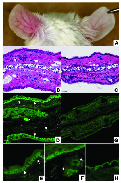
Passive-transfer study using affinity-purified lichen sclerosus IgG in neonatal BALB/c mice. (A) Intradermal injection of affinity-purified lichen sclerosus IgG into BALB/c mouse caused extensive erythematous swelling with telangiectasie (left ear), whereas the control skin injected with either nonimmune human IgG exhibited no evidence of skin inflammation (right ear). This picture was taken at 28 days in a mouse that had received seven separate injections in both ears at days 0, 4, 8, 12, 16, 20, and 24. (B) Light microscopy of the mouse skin injected with affinity-purified lichen sclerosus IgG exhibited a pronounced inflammatory infiltration with edema and dilated blood vessels in the upper-middle dermis. The overlying epidermis showed mild acanthosis. (C) In contrast, the control skin injected with affinity-purified normal human IgG showed only a scanty perivascular infiltration in the dermis. (D) The mouse skin injected with affinity-purified lichen sclerosus antibodies revealed IgG deposition within the lower epidermis (arrows) and surrounding dilated dermal blood vessels (arrowheads). Asterisk indicates the injected site. Higher magnification views revealed an intracellular signal in basal keratinocytes and weaker staining in the suprabasal keratinocytes (E), as well as in the walls of dilated dermal blood vessels (F). (G) In contrast, the control injection with affinity-purified normal human IgG resulted in no immunolabeling. (H) At higher magnification, control injections only resulted in a nonspecific signal in the corneal layers. Scale bar: 50 ∝m.

