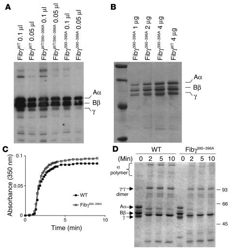
Figure 2.
Characterization of Fibγ390–396A fibrinogen. (A) Western blot of fibrinogen in plasma from WT and mutant mice. (B) Coomassie blue–stained SDS polyacrylamide gel (reducing conditions) showing affinity-purified fibrinogen preparations from WT and Fibγ390–396A mice. (C) Comparative analysis of thrombin-induced fibrin polymerization in plasma from WT and homozygous Fibγ390–396A mice. (D) Analysis of fXIIIa-mediated fibrin cross-linking in reaction mixtures containing either purified WT or γ390–396A fibrinogen. The electrophoretic positions of Aα, Bβ, γ chains are indicated at left along with γ-γ dimer and α polymer cross-linking products.

