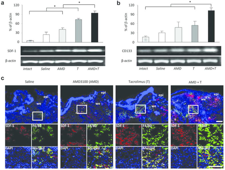
Figure 4. Increased expression of SDF-1 and CD133 in skin wounds of the mice treated with dual drug therapy.

(a) Semi-quantitative RT-PCR analysis of the granulation tissues in wounded skin at 5 days post-injury. The mRNA expression of the attractor molecule SDF-1 was significantly increased in the low-dose Tacrolimus treatment group, and further elevated in the dual treatment group. (b) The stem cell marker CD133 mRNA level was also significantly higher in wounds in the dual treatment group, paralleling SDF-1 gene expression. All data represent means ± SEM. (n = 3). * p < 0.05. (c) Double immunofluorescence staining for SDF-1 and F4/80 (a marker for macrophages) at 5 days after injury. epi, epidermis; we, wound edge. Scale bar: 200 µm.
