
Fig. 1.
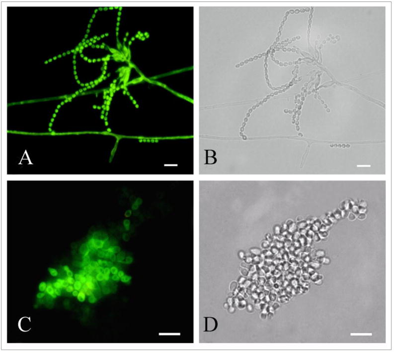
Corresponding immunofluorescence (A,C) and bright field (B,D) microscopy images demonstrating the labeling of mycelial phase (A,B) and yeast cells of P. marneffei by the melanin-binding MAb 8D6. No reactivity was observed when FITC labeled goat-anti mouse IgM alone was used (not shown). Bar, 5 μm.