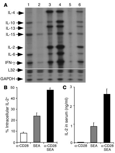
Figure 2.
Anti-CD28 mAb provides costimulatory signals to T cells in vivo. (A) Anti-CD28 mAb increases cytokine mRNA levels in activated T cells in vivo. DO11.10 WT (lanes 1–4) or CD28-deficient (lanes 5 and 6) mice were injected with control mAb plus control peptide (lanes 1 and 5), anti-CD28 mAb plus control peptide (lane 2), control mAb plus OVA peptide (lanes 3 and 6), or anti-CD28 mAb plus OVA peptide (lane 4). One hour after injection, spleens were harvested, and RPA was used to measure cytokine mRNA levels. The lower portion of the gel was exposed to the x-ray film for a much shorter period of time than was the upper portion in order to produce bands of similar intensity for L32 and GAPDH as for other probes. Each band represents one of two mice in the group. Results from one of three representative experiments are shown. (B) Anti-CD28 mAb increases IL-2 expression among activated T cells in vivo. B6 CD80/CD86–deficient mice were injected intravenously with 100 ∝g of anti-CD28 mAb, 10 ∝g/mouse of SEA, or both in combination. Splenocytes were stained for surface expression of CD4 and TCR Vβ3,11 and for intracellular expression of IL-2. The data shown are intracellular IL-2 expression on gated CD4+/TCR Vβ3,11+ (SEA-reactive) cells. (C) Anti-CD28 mAb increases IL-2 production in the serum. Data are shown as averages of two mice in each group with error bars indicating 1 SD. Results represent one of two replicate experiments.

