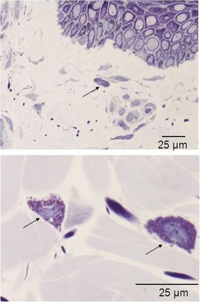
FIG. 29.

Histology for macrophages of in vivo biopsy harvested after three treatments with 35–40% of the full device working power: the arrows highlight the macrophages that have moved from the perivascular niche, and display a slight increase in their count, thus suggesting an active status. Light microscopy, toluidine blue staining, bar 25 μm.
