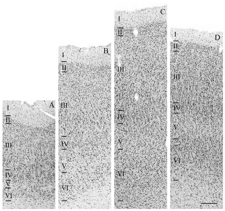
Figure 14.
Example of cytoarchitecture within a temporo-insular fusion extending into white matter in a mountain gorilla (Gorilla beringei beringei), right hemisphere, stained for Nissl substance. Scale bar = 250 μm. Areas shown correspond to those labeled in Fig. 13E and include dysgranular insula (A), medial belt of auditory association cortex (B), the boundary between the medial auditory belt and core of primary auditory cortex (C), and core of primary auditory cortex (D).

