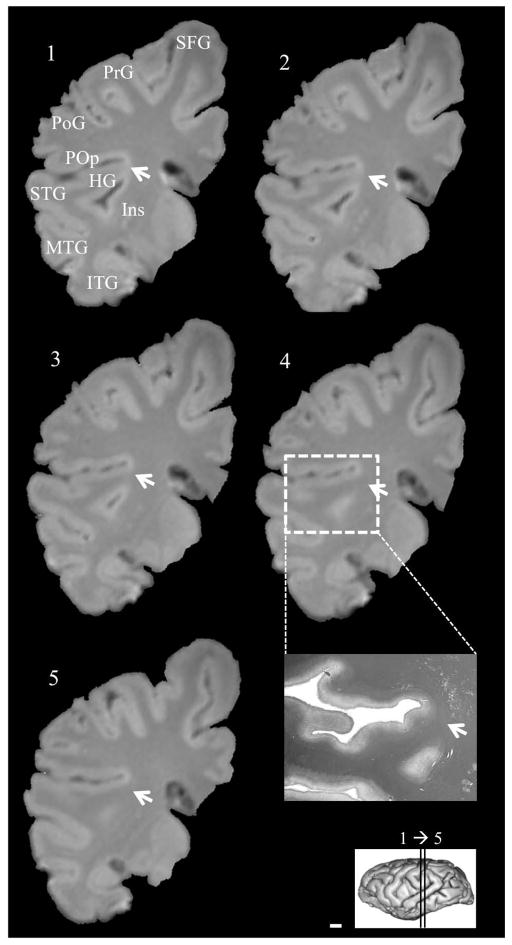
Figure 3.
Serial coronal MRI images of a fusion, including white matter, between posterior temporal lobe and posterior insula in a Grauer’s gorilla (Gorilla beringei graueri), and tissue corresponding to coronal section 4 stained for myelin (inset). Arrows indicate point of fusion. Left hemisphere, anterior to posterior. Numbers indicate level of coronal section. Scale bar = 1 mm. HG: Heschl’s gyrus. Ins: Insula. ITG: Inferior temporal gyrus. MTG: Middle temporal gyrus. PoG: Postcentral gyrus. POp: Parietal operculum. PrG: Precentral gyrus. SFG: Superior frontal gyrus. STG: Superior temporal gyrus.

