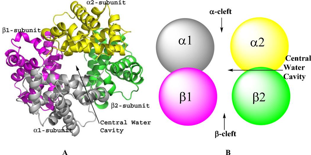
Figure 1.
Quaternary structure of human Hb. α1-subunit (grey), α2-subunit (yellow), β1-subunit (magenta) and β2-subunit (green). (A) Ribbon diagram showing the four Hb subunits arranged around a central water cavity. (B) Spherical illustration of the Hb subunits showing access to the central water cavity via the α-cleft and β-cleft.

