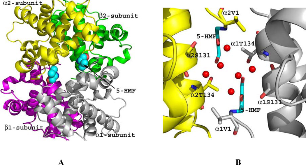
Figure 4.
Crystal structure of quaternary R2 in complex with two molecules of 5-HMF (cyan spheres). Hb subunits are in ribbons (α1-subunit in grey, α2-subunit in yellow, β1-subunit in magenta and β2-subunit in green). (A) Two molecules of 5-HMF bound at the α-cleft in a symmetry-related fashion to the N-terminal αVal1. (B) Close-up view of the bound 5-HMF molecules. Each molecule forms a Schiff-base interaction with the αVal1 nitrogen. The sheath of water molecules (red spheres) form an intricate hydrogen-bond interactions with the bound compounds and the protein to tie the two α-subunits together.

