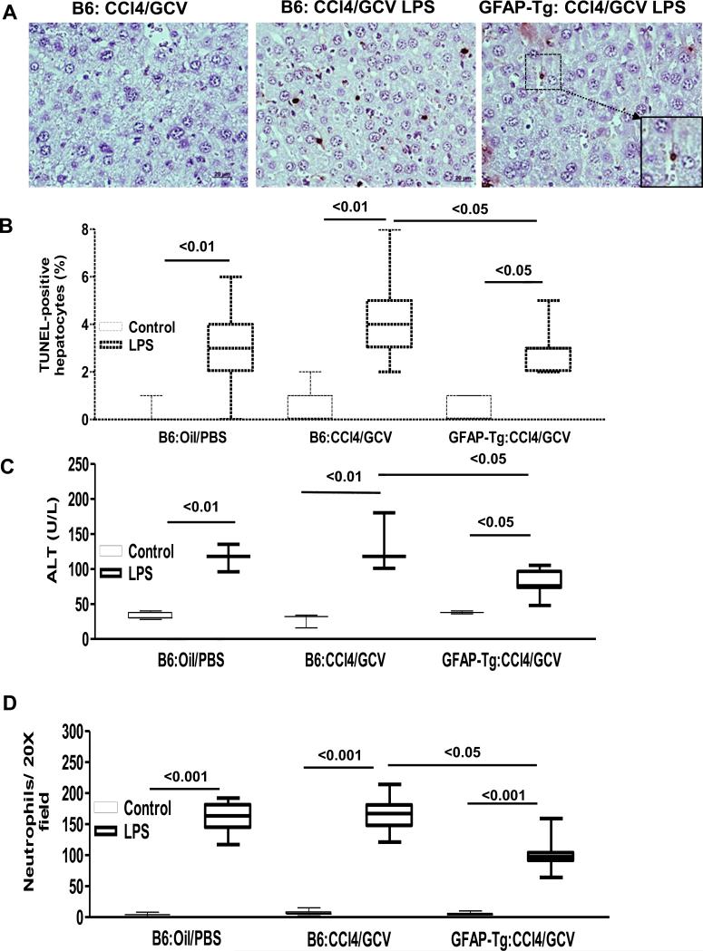
Fig. 3. Hepatic endotoxemic injury in HSC-depleted mice.
WT-B6 and GFAP-Tg mice (n=4-6) received CCl4 (3 injections) followed by its discontinuation and GCV treatment of 10 days, then subjected to LPS treatment. (A) Histopathology and (B) quantification of TUNEL- positive hepatocytes; (C) serum ALT; and (D) neutrophil infiltration. Compared to CCl4/GCV then LPS-treated B6 mice very few apoptotic hepatocytes were observed in the livers of CCl4/GCV then LPS-treated GFAP-Tg mice; inset shows high magnification of the marked area to show apoptotic HSC.

