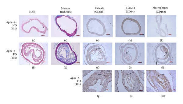
Figure 2.

APOE−/− mice. Advanced atherosclerosis model. ((a), (c)) Histological section of APOE−/− mice fed with ND. ((b), (d)) Histological section of ApoE−/− mice fed with FD. H&E staining and Masson trichrome. (e) Histological section of ApoE−/− mice fed with ND. ((f), (g)) Histological sections of ApoE−/− mice fed with FD. Immunohistochemistry for platelets (CD61), 1/25. (h) Histological section of ApoE−/− mice fed with ND. ((i), (j)) Histological sections of ApoE−/− mice fed with FD. Immunohistochemistry for ICAM-1, 1/25. (k) Histological section of ApoE−/− mice fed with ND. ((l), (m)) Histological sections of ApoE−/− mice fed with FD. Immunohistochemistry for ICAM-1, 1/25. Scale bar: 50 and 200 μm.
