
Figure 3.
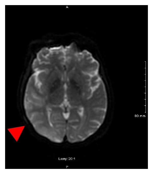
MRI of the brain showing a large area of gyral edema, sulcal effacement, and cortically based diffusion restriction involving the right occipital lobe and right posterior temporal and parietal lobes (indicated by RED arrow).
Official websites use .gov
A
.gov website belongs to an official
government organization in the United States.
Secure .gov websites use HTTPS
A lock (
) or https:// means you've safely
connected to the .gov website. Share sensitive
information only on official, secure websites.
MRI of the brain showing a large area of gyral edema, sulcal effacement, and cortically based diffusion restriction involving the right occipital lobe and right posterior temporal and parietal lobes (indicated by RED arrow).