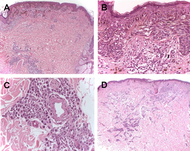
Figure 4.
(A) Superficial spreading melanoma with underlying infiltration of mast cells (H-E, 4 X); (B) Melanocytic atypia with pagetoid spread (H-E, 20 X); (C) Mast cells in the dermis with a particular perivascular arrangement (H-E, 20 X); (D) A rescission of the melanoma showing a scar with the simultaneous presence of a mast cell population.

