Figure 5.

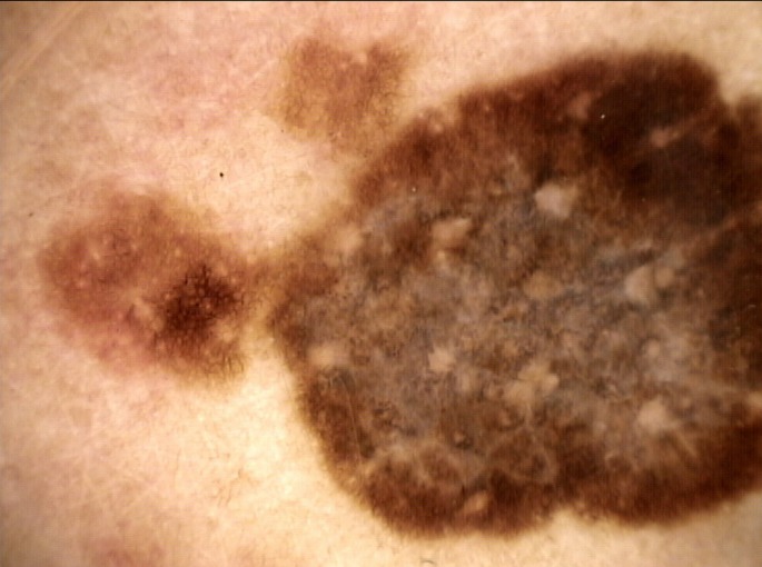
Dermoscopic image of the malignant melanoma that was removed. The lesion showed a broadned-network, radial streaming, inverted network, moth-eaten border, central whitishveil with a scar-like depigmentation and multiple blue-gray dots.

Dermoscopic image of the malignant melanoma that was removed. The lesion showed a broadned-network, radial streaming, inverted network, moth-eaten border, central whitishveil with a scar-like depigmentation and multiple blue-gray dots.