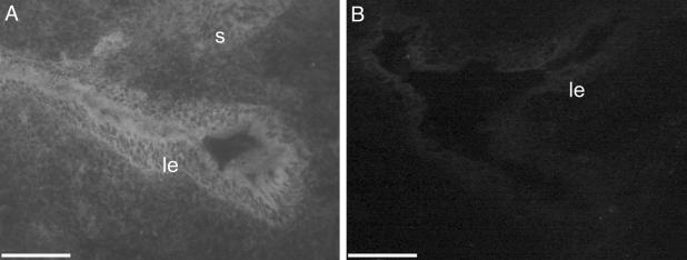
Fig. 2.
Fluorescence micrographs of mouse uteri after intrauterine administration of FITC-MO. (A) Uterine horn treated with control FITC-MO displaying high levels of fluorescence in luminal epithelial compartment and moderate fluorescence in stromal compartment. (B) Uterine horn treated with unlabeled control MO displaying only background fluorescence. (Scale bars = 25 μm.)

