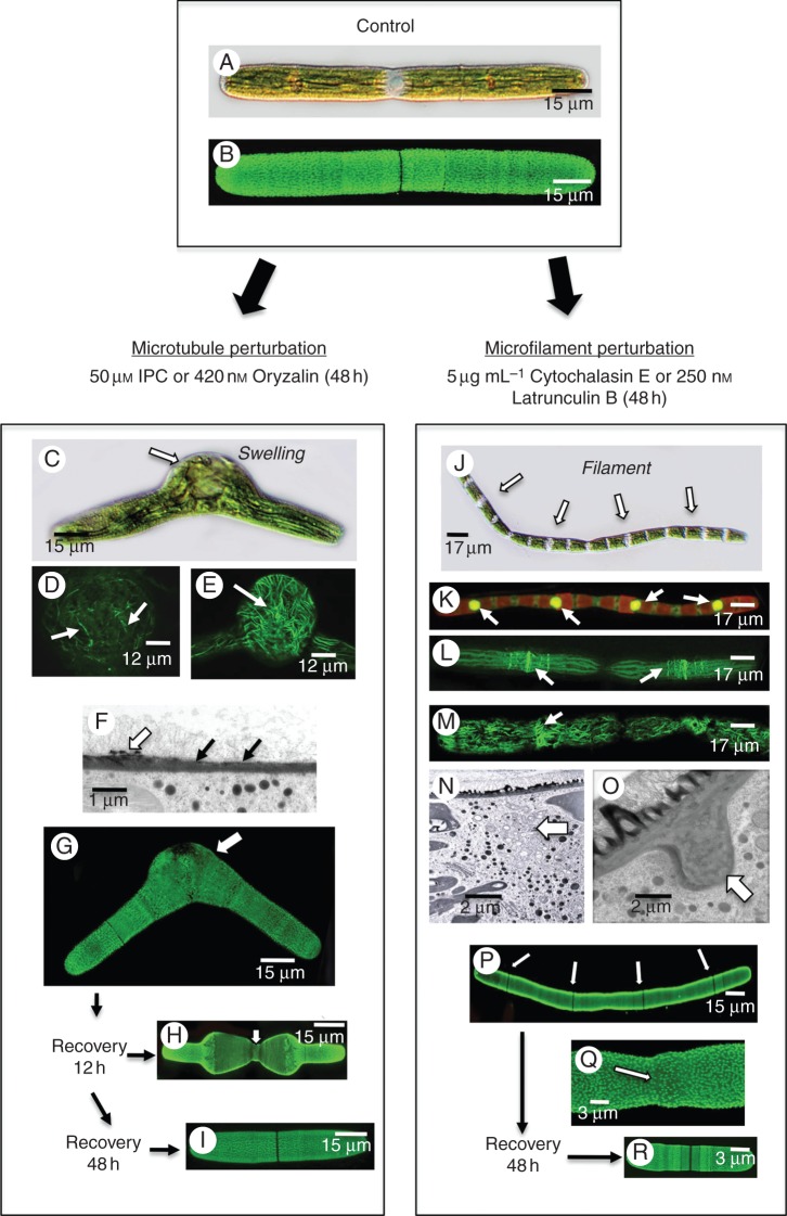
Fig. 5.
Effects of microtubule and actin perturbation agents. (A) DIC image of an untreated cell. (B) LM18 labelling of an untreated cell highlighting the pectin lattice. (C) DIC image of a cell treated with 50 µm IPC for 24 h. Note the distinct swelling at the isthmus. (D) Anti-tubulin labelling of the swollen isthmus region of an IPC-treated cell. No banding is noted and the microtubules are in disarray. (E) Rhodamine phalloidin labelling of the swollen isthmus of an IPC-treated cell. The actin network is disorganized. (F) TEM image of the cell wall at the interface of the regular wall and the isthmus at the swollen isthmus. The cell wall in the isthmus is thinner (arrows) and has no pectin lattice (white arrow). (G) LM18 labelling of an IPC-treated cell. Note the irregular labelling at the swollen isthmus zone. (H) LM18 labelling of a cell washed free of IPC and allowed to recover for 2 h. Note that the cell is thinner at the isthmus. (I) Forty-eight hours after recovery, cells return to normal shape. (J) Pseudofilament formed after cells are treated with cytochalasin E for 48 h. Note the individual cell units (arrows). (K) SYTO9 labelling of pseudofilament demonstrates that each cell unit contains a nucleus. (L) Anti-microtubule labelling of a psuedofilament. Note that the microtubule bands are still present. (M) Rhodamine phalloidin labelling of a pseudofilament showing the disorganization of the cortical actin filaments. No actin filament band is seen near the nucleus. (N) TEM image of the zone between cellular units in a cytochalasin E-treated cell. Note that the cytoplasm consists of a collection of vesicles (arrow). (O) TEM image of the isthmus region of a cytochalasin E-treated cell. Note the large wall plug found at the cell periphery (arrow). (P) LM18 labelling of the pseudofilament showing that little alteration to the pectin lattice (arrows). (Q) LM18 labelling of the isthmus zone between units. Note that the lattice remains intact. (R) Upon 48 h of recovery, cells return to a unicellular phenotype.

