
Fig. 3.
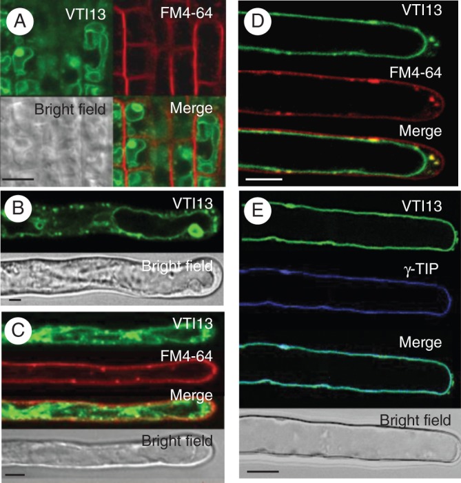
Localization of 35S:GFP–VTI13 in root epidermal cells and root hairs of 5-day-old arabidopsis seedlings. (A) 35S:GFP–VTI13 (green) in root tip epidermal cells highlighting vacuolar ‘bulbs’ and co-stained with FM4-64 (red) for 2–5 min. (B) 35S:GFP–VTI13 in root hairs highlighting vacuolar ‘bulbs’. (C) 35S:GFP–VTI13 (green) in root hairs co-stained with FM4-64 (red) for 2–5 min. (D) 35S:GFP–VTI13 (green) in root hairs co-stained with FM4-64 (red) for 15–20 min. (E) Co-localization of VTI13 (green) and the vacuolar protein YFP-γTIP (purple). The image is a medial section of the z-series through the root hair to show the cross-section view of the cell. Scale bars: (A–D) = 5 μm; (E) = 10 μm.