
Fig. 6.
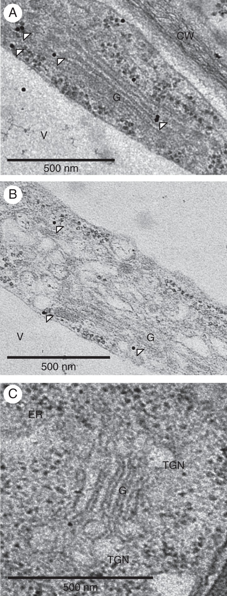
VTI13 localizes to the post-Golgi and trans-Golgi network or early endosomes in arabidopsis root hair cells. GFP–VTI13 was visualized using TEM and α-GFP antibodies conjugated to 10 nm gold particles on root hair sections. (A, B) Longitudinal sections of individual root hair cells expressing 35S:GFP–VTI13 show VTI13 localized to the periphery of the Golgi and post-Golgi compartments. White arrowheads point to representative gold particles labelling VTI13 in these subcellular regions. (C) Longitudinal section of a root hair cell expressing 35S:GFP–VTI13 showed no immunogold labelling when only secondary antibody was used. CW, cell wall; V, vacuole; G, Golgi apparatus. Scale bars: (A–C) = 500 nm.