
Fig. 8.
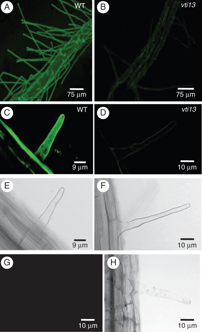
The vti13 mutant seedling root epidermal cells and root hairs exhibit altered cell wall organization. Confocal microscopy analysis of LM15 labelling in wild-type seedlings shows extensive and uniform distribution of xyloglucan in the cell wall of (A) root epidermal cells and (C) root hairs. Conversely, the vti13 mutant showed little to no LM15 labelling of the cell wall of (B) root epidermal cells and (D) root hairs. (E, F, H) Bright-field images of root hairs in (C, D and G), respectively. (G) Secondary antibodies alone did not label the cell wall of wild-type root hairs. All images were taken with the confocal microscope at the same settings to show the difference in labelling at the same setting and antibody concentration. Scale bars: (A, B) = 75 μm; (C, E) = 9 μm; (D, F, G, H) = 10 μm.