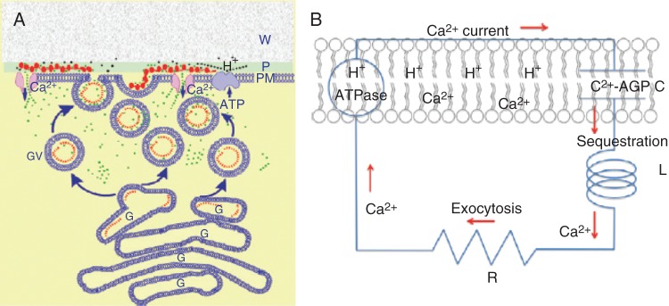
Fig. 5.

The AGP–calcium oscillator. (A) Ca2+ release from periplasmic AGPs. The oscillator generates pulses of Ca2+ (green dots) whose influx coordinates exocytosis and rapid tip growth. This involves a pulse of H+ (black dots) releasing Ca2+ from periplasmic AGPs (red beads) via stretch-activated Ca2+ channels into the cytosol, and then sequestration by exocytotic Golgi vesicles containing AGPs (red dots). Diffusion of the initial H+ pulse to the wall domain restores the periplasmic pH. Ca2+ is recycled by fusion of vesicles with the plasma membrane. Ca2+ bound to periplasmic AGPs is now ready for the next oscillation. W, wall; P, periplasm; PM, plasma membrane; G, Golgi; GV, Golgi exocytotic vesicles. (B) Ca2+ current as a molecular clock: analogous to an electronic series RLC circuit where R is resistance, L is inductance and C is capacitance, with membrane-bound AGPs as the capacitor, C; Ca2+ sequestration as the inductance, L; vesicle exocytosis as the resistor, R, which limits the recycling rate. Hypothetically, C and L largely determine the oscillator frequency and amplitude of the Ca2+ current, ICa. Note the reported high efflux of Cl– as a counterion may maintain electrical neutrality (Cosgrove and Hedrich, 1991; Zonia et al., 2002). [Reprinted from Lamport and Varnai (2013).]
