Figure 1.

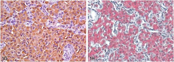
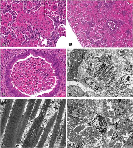
Eosinophilic crystalline pneumonia in transgenic mice. A, Pulmonary alveolar spaces are markedly distended with numerous large epithelioid macrophages with brightly eosinophilic granular to amorphous cytoplasm containing fine acicular crystalline material. Occasional large binucleate and multinucleate forms were also observed (inset). Hematoxylin and eosin (H&E). B, Accompanying the marked alveolar cellular infiltrates in these mice, there was variable alveolar wall thickening with mononuclear cell infiltrates and peribronchiolar fibrosis. (H&E). C, Variable bronchiolar epithelial cell hypertrophy associated with intraluminal infiltrates consisting of aggregates of viable and degenerating macrophages and neutrophils. (H&E). D, Alveolar macrophages were markedly enlarged and distended with electron dense, needle-shaped cytoplasmic inclusions (crystalloid arrays). Transmission electron micrograph. E, Higher magnification of crystalloid arrays seen in (D). Crystalloid arrays were composed of linear stacks of granular to amorphous membrane-bound electron-dense material. Transmission electron micrograph. F, Similar electron dense crystalloid inclusions were observed in Kupffer cells lining the hepatic sinusoids. Transmission electron micrograph. G, Labeling with antibodies against Chi3l3 protein showed moderately to markedly immunoreactive macrophages with intracytoplasmic crystalline material. H, Labeling with Luna histochemical stain showed positively stained macrophages with intracytoplasmic crystalline material.

