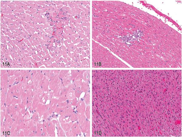
Figure 11.
Cardiovascular lesions in 10- to 12-week-old Sprague-Dawley rats. A, A well-circumscribed focus of hyaline cardiomyocyte eosinophilia with a few marginating neutrophilic inflammatory cells within the myocardium. B, A focal accumulation of mixed mononuclear inflammatory cells that have responded to and replaced a focus of cardiomyocyte necrosis. C, An area of myocardium where a number of individual cardiomyocytes are distorted by clear vacuoles that are irregular in shape and size and might be considered to expand the cellular cytoplasm. D, An area of myocardium with clear vacuoles that are present in much greater number than that compared to (C) and are more consistent in size and shape but appear to have less effect on the overall morphology of the cells (H&E).

