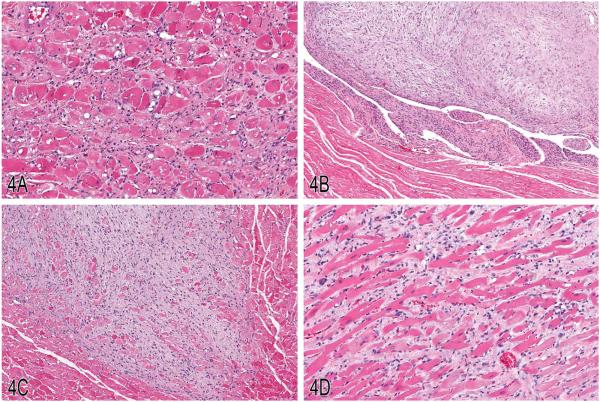
Figure 4.
Examples of “rodent progressive cardiomyopathy” (RPM), schwannoma, and cardiomyopathy in rats. A, An example of RPM characterized by a locally extensive region of cardiomyocyte necrosis and replacement by fibrosis and mononuclear infiltrates, including pigment-laden macrophages. B, A lesion diagnosed as “schwannoma” with a mildly anisocytotic and anisokaryotic subendocardial spindle cell proliferation, spanning the endocardium from the apical endocardium to the basilar endocardium. C, Example of an intramural schwannoma with interwoven bundles of neoplastic spindle cells replacing and displacing nascent cardiomyocytes. D, A lesion diagnosed as “cardiomyopathy” characterized by a regionally extensive spindle cell proliferation that dissects and displaces cardiomyocytes. (H&E).

