
Figure 3. Purkinje cell degeneration in SCA7 involves Bergmann glia dysfunction.
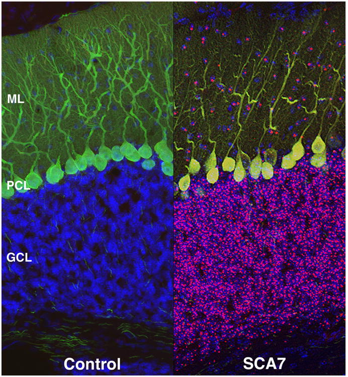

a) Non cell-autonomous SCA7 Purkinje cell degeneration. Confocal microscopy analysis of cerebellar sections from a PrP-SCA7-c92Q mouse (SCA7) and from a non-transgenic littermate (Control). Immunostaining with anti-ataxin-7 antibody (magenta), calbindin antibody (green), and DAPI (blue) yields normally oriented Purkinje cells with extensive dendritic arborization in the ‘Control’ mice. However, SCA7 transgenic mice display decreased dendritic arborization and displacement of Purkinje cell bodies, indicative of degeneration. While neurons in the granule cell layer (GCL) and the molecular layer (ML) contain aggregates of ataxin-7 protein, mutant ataxin-7 protein is not expressed in degenerating Purkinje cells. Degeneration of Purkinje cells without expression of mutant ataxin-7 protein is a so-called “non cell-autonomous” process. Adapted from Garden et al., 2002, J Neurosci 22, 4897. Used with permission.
b) Bergmann glia expression of mutant ataxin-7 protein is sufficient to produce Purkinje cell degeneration. Immunohistochemistry analysis of cerebellar sections from non-transgenic (NT), Gfa2-SCA7-10Q (10Q), and Gfa2-SCA7-92Q (92Q) mice was performed with anti-ataxin-7 (green) and anti-calbindin (red) antibodies. NT mice have normal cerebellar cytoarchitecture, as do 10Q mice. Reduced molecular layer (ML) thickness, misaligned Purkinje cells in the Purkinje cell layer (PCL), and tortuous Purkinje cell dendrites characterize the 92Q cerebellum. Note intense, punctate ataxin-7 immunostaining in the 92Q PCL (arrows), unlike the diffuse ataxin-7 staining observed in the 10Q PCL (arrows). GCL = granule cell layer. Adapted from Custer et al., 2006, Nat Neurosci 9, 1302. Used with permission.