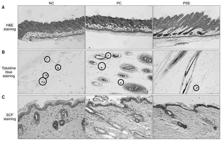
Fig. 3.
Representative histological observation, comparison of number of mast cells, and immunohistochemistry of SCF antigens in an alopecia model of C57BL/6 mice treated with topical test compounds for 3 weeks. Shape and depth of H&E stained follicles (A), toluidine blue-stained sections showing mast cell count (B), and immunohistochemical staining of SCF in the follicles, mast cells, and skin (C). NC, dimethyl sulfoxide (DMSO); PC, minoxidil; PSE, Platycarya strobilacea S. et Z. extract with DMSO.

