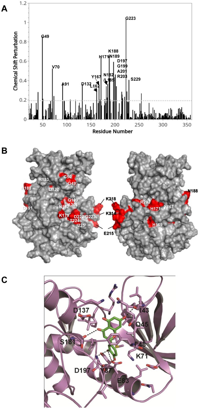
Figure 3. NMR titration assay and in silico modeling for interaction of luteolin with VRK1.
(A), NMR titration experiments were performed. Spectrum of chemical shift perturbations versus amino acid residues of the VRK1 protein after binding of luteolin. (B), Mapping of chemical shift perturbations on the VRK1 protein. Most of the perturbed residues (shown in red) are located close to the catalytic domain of VRK1. (C), in silico modeling of the binding mode of luteolin to the VRK1 protein. Luteolin is predicted to fit in the vicinity of the G-loop, catalytic site, and α-C lobe.

