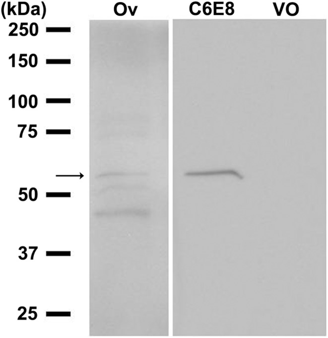
Figure 6. Detection of S. invicta sNPF receptor in cell membranes prepared from the SisNPFR-C6E8 cell line by western blot analysis.

Numbers in the left indicate the marker’s protein mass (kDa). Lane 1, the membrane fraction of the fire ant mated queen ovary (Ov) as a positive control; lane 2, the membrane fraction of SisNPFR-C6E8 (C6E8) and lane 3, the membrane fraction of the vector-only (VO) (pcDNA3.1(-)) transfected CHO-K1 cells probed with the specific anti-sNPF receptor anti-peptide antibody [44]. Membrane protein (50 µg) was loaded in each lane. The antibody detects the 55 kDa receptor protein in lanes 1 and 2, as expected.
