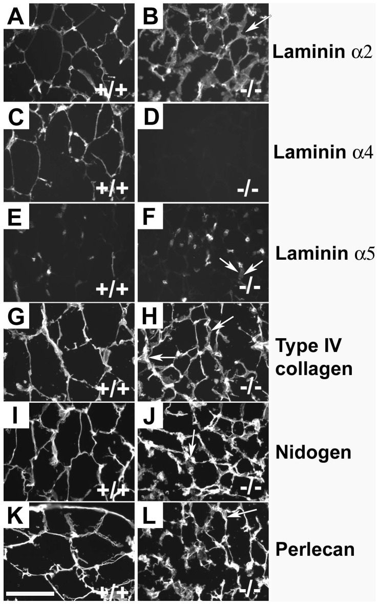
Figure 1. Immunostaining of WAT.
The capillaries and BMs of adipocytes of wild-type control and Lama4Lama4−/− mice were positive with antibodies against laminin α2 (A, B). Laminin α4 antibodies stained both adipocytic BMs and capillaries in wild-type controls (C), while no staining was seen in mutants (D). The laminin α5 antibody (E, F) stained capillaries in Lama4−/− and wild-type controls, but not the adipocytic BM. Antibodies against type IV collagen (G; H), nidogen (I, J) and perlecan (K, L) all stained both capillaries and adipocytic BMs. Laminin α1 staining was completely negative (not shown). Arrows indicate a few locations where the adipocytic BM of the Lama4−/− mice appeared slightly thickened. Bar 100 µm.

