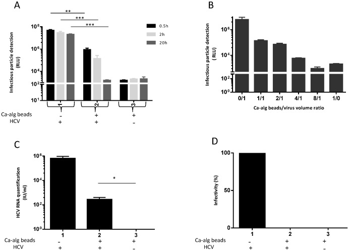
Figure 4. Protective property of Ca-alg beads against HCV infection is dependent on bead/virus volume ratio concentration and time of incubation.
Ca-alg beads devoid of cells were produced. (A) 1200 µL of Ca-alg beads were transferred in tissue culture 6-well plates with 1 mL of complete DMEM medium added on 3D moving plates. After three incubation times (0.5, 2 and 20 h) at room temperature of JHF1-RLuc virus with a Ca-alg bead/virus volume ratio of 4/1, the supernatant was recovered and incubated for 4 h with HuH-7 cell 2D cultures. The detection of infectious particles was assessed by luciferase assay on infected cells at 72 h post-infection. Results are expressed as RLU and are reported as the means ± S.D. of three independent experiments. (B) The same experiment was performed with different Ca-alg bead/virus volume ratios (0/1 to 8/1) for 20 h. As previously described, the supernatant was recovered and incubated for 4 h with HuH-7 cell 2D cultures. Luciferase assays were performed on the infected cells at 72 h post-infection. The Ca-alg beads without the JFH1-RLuc virus were used as controls (Ctrl). Results are expressed as RLU and are reported as the means ± S.D. of three independent experiments. (C) After 20 h post-incubation of JFH1 virus in a Ca-alg bead/virus volume ratio of 4/1, the Ca-alg beads were washed, degelified by lyase treatment. Viral titers of the supernatants were determined by FFU assay. Results are converted into a percentage of infectivity. (D) Under the same incubation conditions as C), the amount of HCV RNA was also quantified in the supernatants (condition without beads) and from the Ca-alg bead products after lyase treatment (condition with beads) by RT-qPCR. Results are expressed as HCV RNA IU/mL and are reported as the mean ± S.D. of triplicate measurements. *P<0.05, **P<0.001, ***P<0.0001.

