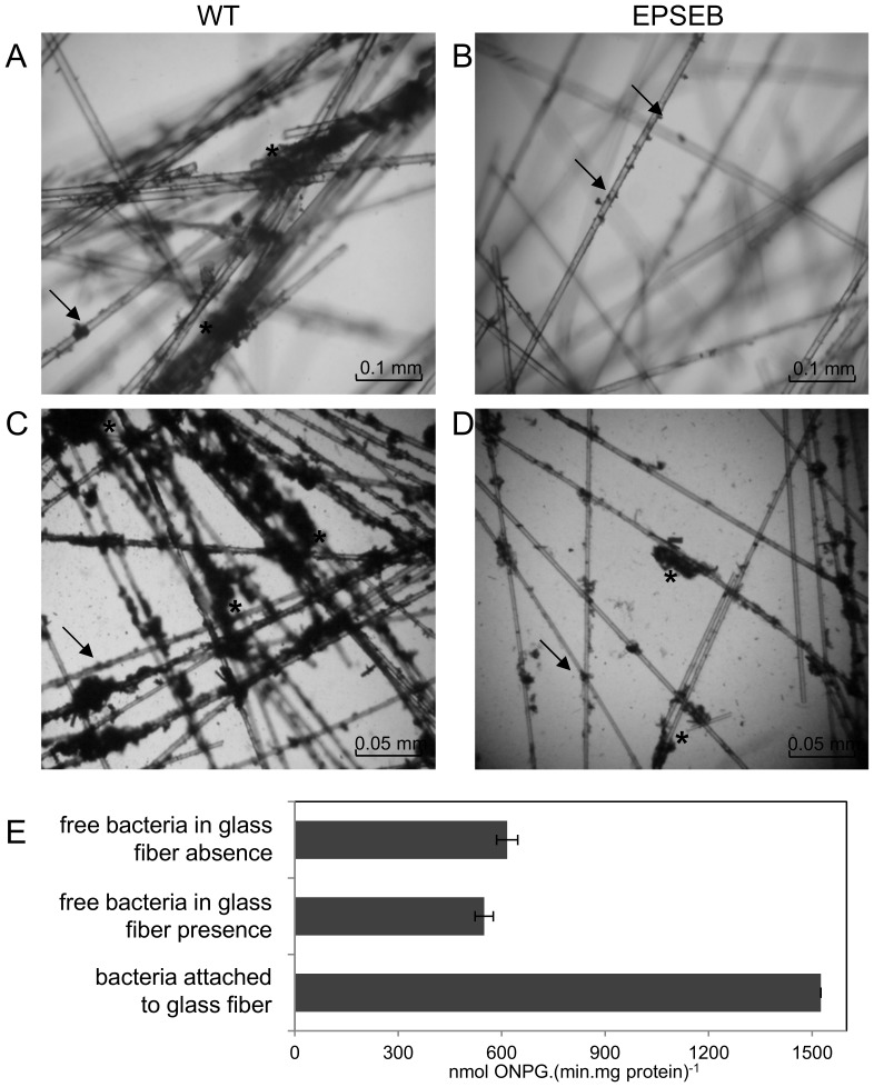
Figure 2. H. seropedicae biofilm formation on glass fiber.
Light microscopy was performed with H. seropedicae SmR1 and EPSEB (epsB mutant) grown in the presence of glass fiber for 12 hours, without (A,B) and with (C,D) addition of purified wild-type EPS (100 µg.mL−1). Arrows indicate attached bacteria. Asterisks indicate mature biofilm colonies. For biofilm expression analyses (E), H. seropedicae MHS-01 cells were grown for 12 h in the presence or absence of glass fiber, the free living bacteria were directly used and biofilm bacteria were recovered from glass fiber by vortex. β-galactosidase activity was determined, standardized by total protein concentration, and expressed as nmol ONP.(min.mg protein) −1± standard deviation. Different letters indicate significant differences (p<0.01, Duncan multiple range test) in epsG expression between the tested conditions.

