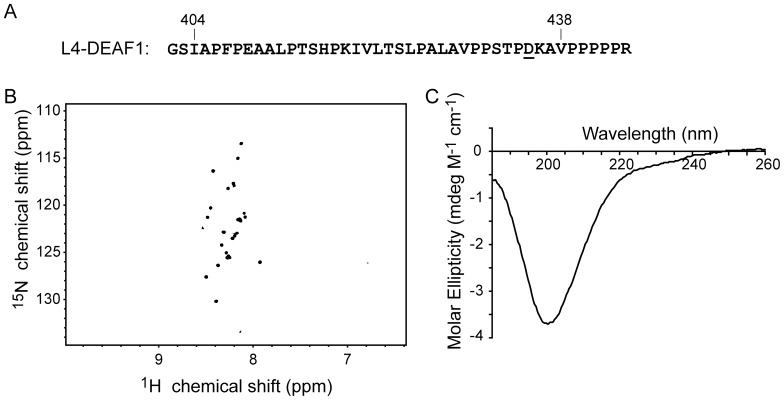
Figure 2. The LMO4-binding domain from DEAF1 is disordered in solution.
(A) The sequence of L4-DEAF1 includes residues 404–438 of DEAF1 (bold), a T435D point mutation (underlined) and a polyproline C-terminal tail (PPPPPR). The two N-terminal residues (GS) are an artefact of the plasmid and remain after treatment with thrombin. (B) 15N-HSQC spectrum of L4-DEAF1 (160 µM) was recorded in 20 mM sodium acetate at pH 5.0 and 35 mM NaCl at 298 K on a 600 MHz spectrometer equipped with a TCI-cryogenic probehead. (C) The far-UV CD spectrum of L4-DEAF1 (40 µM) dissolved in 20 mM Tris-acetate at pH 8.0 and 50 mM NaF.

