
Figure 1. Depletion of XIAP leads to enhanced activation of the MEKK2/3-MEK5-ERK5 pathway.
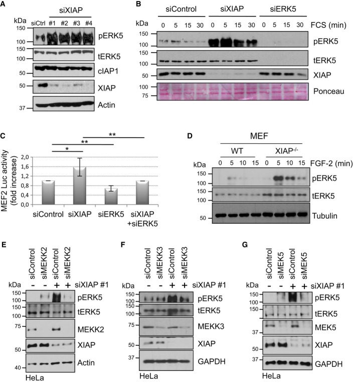
A Depletion of XIAP increases the basal phosphorylation of ERK5. HeLa cells were transiently transfected with Control or XIAP siRNAs. Total lysates were analyzed by Western blotting with various antibodies.
B Depletion of XIAP enhances the strength of ERK5 activation. HeLa-MEF2 cells were transiently transfected with siControl or siXIAP or siERK5 and then stimulated with 10% FCS after serum starvation. Total lysates were analyzed by Western blotting.
C Depletion of XIAP enhances MEF2 transcriptional activity. HeLa cells stably expressing the MEF2-luciferase (luc) reporter gene were transiently transfected with siRNAs against XIAP, ERK5, or both. Cells were then lysed and the luciferase activity was measured and normalized to the Control activity as mentioned in the methods section (shown is the quantification of five independent experiments with *P < 0.05 and **P < 0.01, Student's t-test).
D Loss of XIAP in MEFs also enhances ERK5 phosphorylation. WT, and XIAP−/− MEFs were stimulated with 25 ng/ml of FGF-2. Total lysates were analyzed by Western blotting.
E–G XIAP-mediated effect on ERK5 phosphorylation is dependent of MEKK2, MEKK3, and MEK5. HeLa cells were co-transfected with siRNAs against XIAP and/or MEKK2 (E), MEKK3 (F), or MEK5 (G). Total lysates were analyzed by Western blotting as indicated.
Source data are available online for this figure.