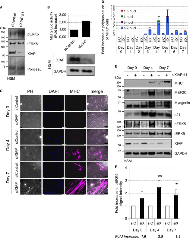
Figure 6. Depletion of XIAP enhances myoblasts differentiation.
A Depletion of XIAP leads to an increase in ERK5 phosphorylation in undifferentiated myoblasts. Undifferentiated HSM were transiently transfected with siControl or siXIAP.
B Depletion of XIAP leads to an increase in ERK5 activation in undifferentiated myoblasts. Undifferentiated HSM cells stably expressing the MEF2-luciferase (Luc) reporter gene were transiently transfected with siControl or siXIAP. Part of the lysates was used for Western blot detection and another part to measure the luciferase activity (shown is the fold increase of MEF2 activity upon XIAP knockdown).
C, D Depletion of XIAP leads to an increase of myoblasts differentiation. HSM were transiently transfected with siControl or siXIAP, and differentiation was started the next day. Transfection was performed every 2 days, and media were changed every following day. Coverslips were fixed and stained everyday as described in methods (shown in (C) are data from a representative experiment from day 0, day 4, and day 7). Polynucleation of MHC positive cells was quantified from four independent experiments, and the average fold increase is shown in (D).
E, F Depletion of XIAP leads to an increase in ERK5 phosphorylation and expression of differentiation markers in myoblasts. HSM were transfected and differentiated as described for (C). Cells were lysed, run on SDS–PAGE, and blotted for various differentiation markers (E). Increase in ERK5 phosphorylation was quantified from three independent experiments, and the average fold increase is shown in (F) (Student's t-test, *P < 0.05, **P < 0.01).
Source data are available online for this figure.

