
Figure 7. PPS-negative auto-regulation.
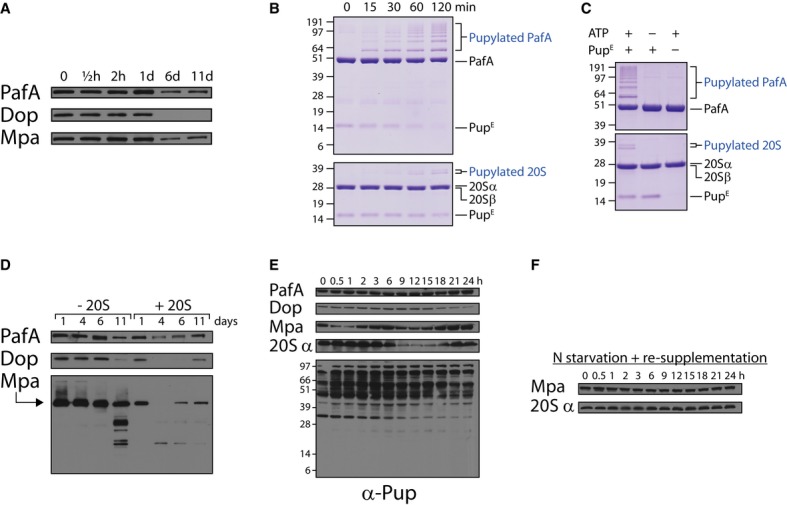
- As in Fig6A, except that antibodies against the indicated proteins were used in the Western blot analysis.
- 20 μM PupE were incubated in pupylation buffer at 30°C together with 5 μM PafA (upper panel) or with 1 μM PafA and 5 μM 20S (lower panel). The first sample was removed (t = 0), ATP (2 mM) was added to start the reactions, and additional samples were removed at the indicated time points for SDS–PAGE followed by Coomassie Brilliant Blue staining.
- Reactions were mixed as in (B) with or without ATP or PupE, as indicated. Following a 2-h incubation at 30°C, aliquots were collected and subjected to SDS–PAGE followed by Coomassie Brilliant Blue staining.
- As in (A), except that a ΔprcSBA mutant was used that expresses from a chromosomally integrated plasmid either the prcS gene (designated -20S) or the prcSBA operon (designated +20S).
- Western blot analysis using antibodies against the indicated proteins was performed on samples collected at the indicated time points during a nitrogen starvation experiment that was performed as described in Fig6A.
- As in (D), except nitrogen source was re-supplemented following an hour of starvation.