
Figure 2. Cryo-EM of encapsulin nanocompartments and EncA shells.
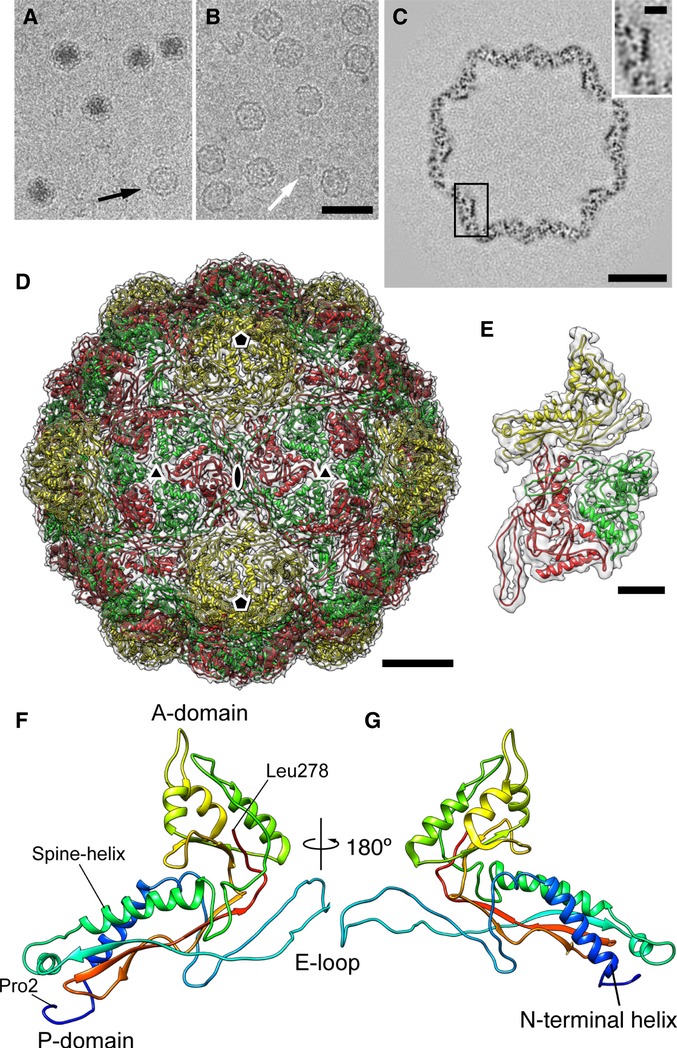
A, B Cryo-micrographs of (A) native nanocompartments and (B) recombinantly expressed EncA capsids.
C Section through a reconstruction of the EncA capsid. An enlargement of the boxed area showing a longitudinal section through an α-helix is inset (upper right).
D Atomic model of the T = 3 EncA capsid. The three quasi-equivalent subunits are colored yellow, green, and red. Symmetry axes are marked.
E The 3-subunit asymmetric unit fitted into electron density (transparent).
F, G Atomic model of EncA, rainbow-colored with the N-terminus in blue, and the C-terminus in red.
Data information: Scale bars: (A, B) 50 nm; (C) 10 nm; (D) 5 nm; (E, C insert) 2 nm.