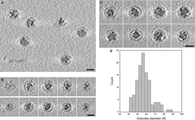
Figure 4. Cryo-ET of encapsulin nanocompartments with electron-dense cores.
- Tomographic slice showing several native Myxococcus xanthus encapsulin nanocompartments.
- Tomographic slices through two nanocompartments (top and bottom rows).
- Gallery of tomographic central sections of eight nanocompartments.
- Histogram of the diameters of electron-dense granules in the nanocompartment cores.
Data information: Scale bars, 25 nm.

