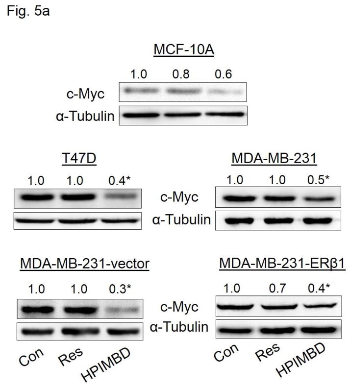
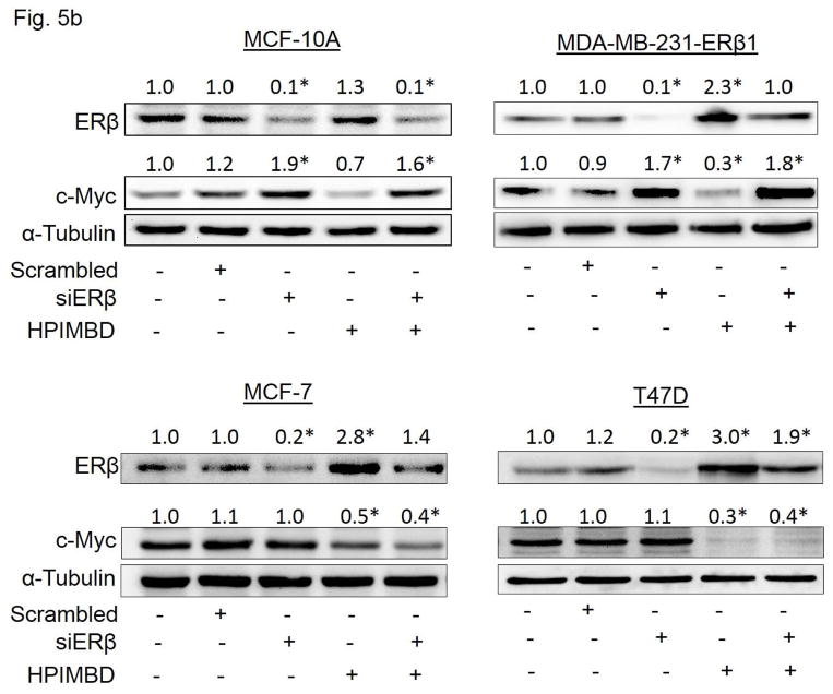
Fig. 5. HPIMBD inhibits the expression of oncogene c-Myc in breast cancer cell lines.
a) Non-neoplastic breast epithelial cell line MCF-10A and breast cancer cell lines T47D, MDA-MB-231, vector-transfected and ERβ1-transfected MDA-MB-231 cells were treated with vehicle (DMSO), 50 μM Res or HPIMBD. Proteins were isolated and western blot analyses were performed. Intensities of the bands were quantified and normalized to α-tubulin. Fold changes in c-Myc protein expression (Mean + SEM) treated with Res or HPIMBD compared to vehicle-treated controls were calculated from four individual experiments and are given at the top of each blot.
b) MCF10A, ERβ1-transfected MDA-MB-231, MCF-7 and T47D cells were transfected with either 1 nmol/l of scrambled small interfering RNA or siERβ for 48 hours, and subsequently treated with 50 μM HPIMBD for 12 hours (ERβ1-transfected MDA-MB-231), 24 hours (MCF-10A) or 48 hours (MCF-7 and T47D). The treatment time points are based on maximal induction time of ERβ for respective cell lines following HPIMBD treatment. Proteins were isolated and western blot analyses were performed. Intensities of the bands were quantified and normalized to α-tubulin. Fold changes in ERβ or c-Myc protein expression (Mean + SEM) treated with scrambled, siERβ or HPIMBD compared to vehicle-controls were calculated from four individual experiments and are given at the top of each blot.
(*) indicates a P value <0.05 compared to controls.


