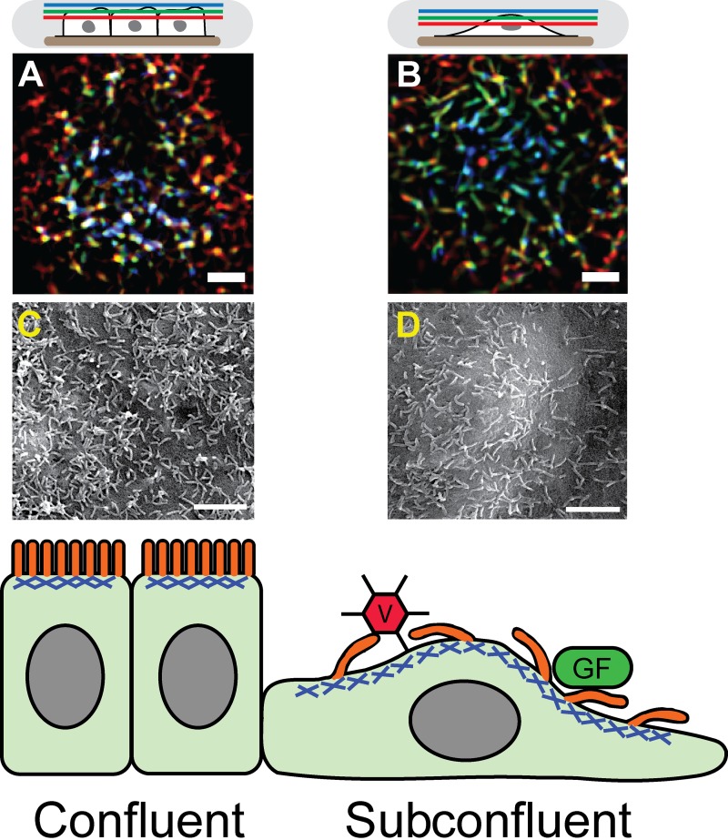
Figure 1.
Transition to subconfluence in epithelial cells alters microvilli morphology and activity. Confluent cell microvilli are more vertically oriented than those in subconfluent cells. (A and B) Color-coded height projections of confocal images of GFP-LifeAct (labels actin) in the apical microvilli of epithelial cells either in confluent (A) or subconfluent (B) conditions. As shown in the schematic on top, blue colors indicate distal z planes, green intermediate z planes, and red apical membrane proximal z planes, so that combined colors represent a vertical structure found in multiple z planes, with white showing structures spanning all three planes. Microvilli are more sparsely spaced on subconfluent cells than on confluent cells. (C and D) Scanning electron micrographs of epithelial cells either confluent (C) or subconfluent (D) to show microvilli morphology and density. A–D have been reproduced from Klingner et al. (2014). See the article for further details. Bars, 2 µm. The bottom panel shows a diagram of epithelial cells in the confluent (left) or subconfluent (right) wound edge. Microvilli, shown in orange, are bound into the apical acto-myosin cortex, shown in blue. In subconfluent cells, the longer, more motile microvilli may enable enhanced binding and uptake of virus (V) or growth factors (GF).

