Figure 3.
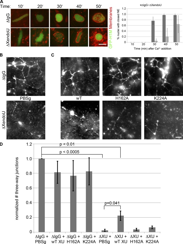
XendoU plays a role in nuclear envelope formation and proper ER morphology. (A) Demembranated X. laevis sperm nuclei were incubated in mock-depleted (IgG) or XendoU-depleted CSF in the presence of GFP-Histone H1 and Vybrant DiI membrane dye. 10–15 random fields were taken from live images every 10 min and the percent nuclei with closed nuclear envelopes was counted. n = 3. (B) IgG mock-depleted (top) or XendoU-depleted (bottom) CSF was incubated at room temperature in the presence of 1 mM CaCl2 and 1× PBS + 15% glycerol (PBSg, protein storage buffer) for 60 min followed by staining of membranes by octadecyl rhodamine and imaged live. (C) Mock-depleted (top) or XendoU-depleted (bottom) extracts were incubated with recombinant proteins (wild-type [left], H162A [middle], or K224A [right]) on ice for 30 min followed by addition of 1 mM CaCl2 and incubated for 60 min at room temperature. Membranes were stained and imaged as above. (D) 10–12 randomly selected fields were taken for each condition in B and C in three separate egg extracts. Three-way junctions between ER tubules were counted for each field to assess network formation. Statistical comparison of IgG + PBSg to experimental samples was performed using a one-sample t test. Comparison of XendoU-depleted to rescued extract was performed using an unpaired Student’s t test. Error bars indicate SD. Bars, 10 µm.

