Figure 4.
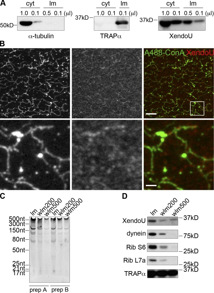
A subpopulation of XendoU localizes to ER membranes. (A) Cytosol (cyt) and light membrane (lm) preps (∼20×) were isolated as described in the Materials and methods. Western blots were performed on each fraction for α-tubulin, TRAPα, and XendoU. (B) ER networks were formed in flow cells and fixed with paraformaldehyde and glutaraldehyde. Immunofluorescence for XendoU was performed with αXendoU antibody and α-rabbit Cy3 secondary antibody (red). Alexa fluor 488 Concanavalin A (conA) was added in with secondary antibodies to detect glycoproteins and the ER (green). The boxed region is magnified 5× in the lower panels. Bars: (top) 10 µm; (bottom) 2 µm. (C) Light membranes (lm) were washed in buffers containing 200 mM (wlm 200) or 500 mM (wlm 500) KCl. Membranes were washed twice and resuspended in the same original volume. RNA was isolated from each membrane prep (prep A and B), run on denaturing acrylamide gels, and stained with SYBR Green II. (D) Western blots for XendoU, dynein, ribosomal protein S6 (Rib S6), ribosomal protein L7a (Rib L7a), and TRAPα were performed on light membranes (lm) and membranes washed in 200 mM KCl (wlm 200) or 500 mM KCl (wlm 500). TRAPα serves as a loading and wash control.

