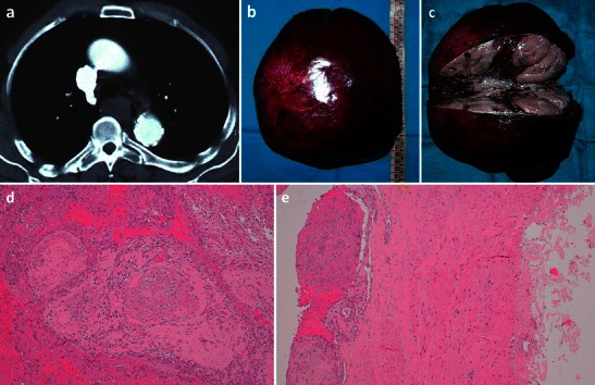
Fig. 10.

Masson’s tumour of the azygos vein. An 80-year-old man who presented with significant dysphagia and hemoptysis, in the absence of any oesophageal diseases. CT revealed a hypodense lesion measuring about 3.5 cm (a) in the azygo-oesophageal recess. With videothoracoscopy the solid lesion (b) of the distal third of the azygos vein, close to the its distal cervical branches, was resected. Macroscopic examination (c) showed an aneurysmatic dilatation of the azygos segment with a luminal thrombus adherent to the vein wall. Histological analysis shows multiple foci of intravascular papillary endothelial hyperplasia were present (d–e)
