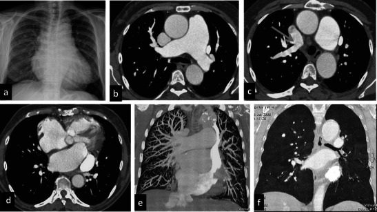
Fig. 3.

Azygos agenesis. A 64-year-old female with a prior mitral valve cardiac substitution. She underwent the PE protocol because of severe dyspnoea and hypoxaemia. Chest X-ray (a) shows increased pulmonary vasculature and mild dilatation of the upper mediastinum particularly on the left side and enlargement of the mediastinal pedicle. Contrast-enhanced CT shows severe enlargement of the pulmonary artery (b) and anomalous venous return with a right upper lobe pulmonary vein draining into the superior vena cava (c). On the left side a persistent left superior vena cava is seen draining into the coronary sinus (d), which is enlarged, too. Marked dilatation of the right atrium and ventricle can be seen. Azygos and hemiazygos veins are absent, suggesting agenesis. Coronal MIP and MinIP reconstructions (e–f) show an increase of the pulmonary vasculature (e) and moderate mosaic perfusion (f) related to severe pulmonary hypertension
