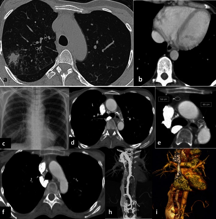
Fig. 6.

Atypical mycobacterial infection: induced fibrosis mediastinitis. A 22-year-old female, affected by celiac disease, underwent CT for fever and cough. It showed a consolidation and tree-in-bud pattern in the right upper lobe (a). The heart and azygos vein were normal (arrow) in size (b) . Bronchoscopy identified an atypical mycobacterial infection. One year later, she underwent a chest X-ray, which showed an enlargement of the superior mediastinum even though the heart size was normal (c). Contrast-enhanced CT scan showed a significant enlargement of the azygos vein, not present in the prior exam (d–f), associated with an increased density of the mediastinal tissue (e). A retrograde, early opacification of the azygous vein can be seen in the axial (d–f) and coronal MPR (h) and VR (f). In the coronal view, the inferior vena cava was enlarged as well. The patient had a fibrosing mediastinitis caused by an atypical mycobacterial infection that induced a reduced calibre of the superior vena cava in the infra-azygos region and retrograde flow in the azygos system
