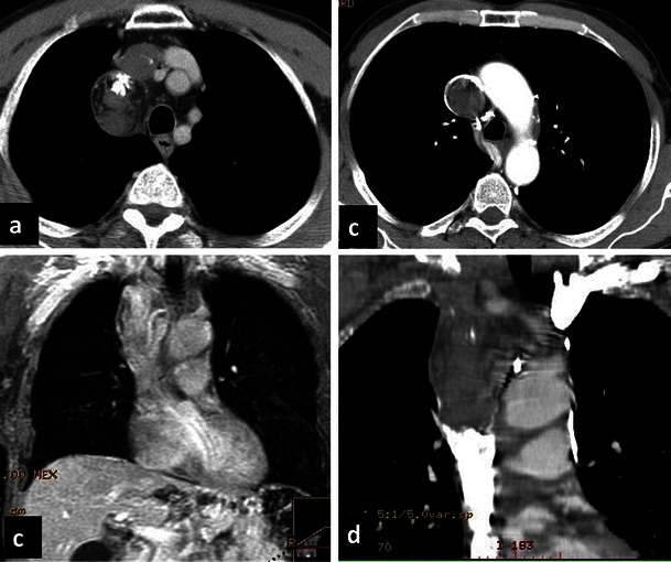
Fig. 9.

Teratoma. Non-contrast and contrast-enhanced CT (a, b) shows a rounded nodule, with some intralesional fatty spots and calcifications in the right paratracheal space consistent with teratoma. The benign lesion induced extrinsic compression of the superior vena cava
