Fig. 18.

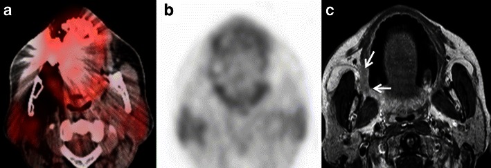
a Axial PET/CT image demonstrates extensive streak artefacts from right-sided dental implants in a patient with clinically proven SCCof the right retromolar trigone. The known lesion in the right retromolar trigone is completely obscured by the streak artefacts. b Corresponding axial FDG-PET image depicts no uptake in the region of the right retromolar trigone thereby yielding a false-negative result. c Corresponding axial contrast-enhanced T1W MR image detects the infiltrative mass in the right retromolar trigone (white arrows). The extent of the lesion as seen on MRI was afterwards confirmed surgically. MRI is less affected by dental artefacts as compared to CT
