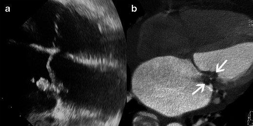
Fig. 1.

Results of echocardiography and MSCT studies in cases of mitral valve IE. Images show four-chamber views in the TEE study (a) and four-chamber view MSCT acquisitions with MPR reconstruction (b). Both TEE and MSCT show a large vegetation (white arrow) and destruction of the mitral valve with substantial dilatation of the left atrium and pericardial effusion
