Fig. 11.

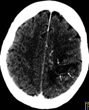
Contrast-enhanced cerebral CT shows a brain abscess as a hypodense, oval shaped lesion with peripheral enhancement (white arrows) and surrounding oedema in the left parietal region

Contrast-enhanced cerebral CT shows a brain abscess as a hypodense, oval shaped lesion with peripheral enhancement (white arrows) and surrounding oedema in the left parietal region