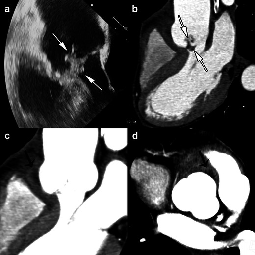
Fig. 4.

Results of echocardiography and MSCT studies in a case of aortic valve IE. The TEE study, 120-degree long-axis view (a) and MSCT acquisition with MPR reconstructions in the LVOT view (b), show a huge vegetation on the aortic valve (white arrows). MSCT acquisitions with MPR in the LVOT view (c) on the level of aortic annulus (d) are displayed in a narrowed imaging window, suitable for analysing para-valvular tissue, and show an abscess located in the anterior part of the aortic root (black arrows)
