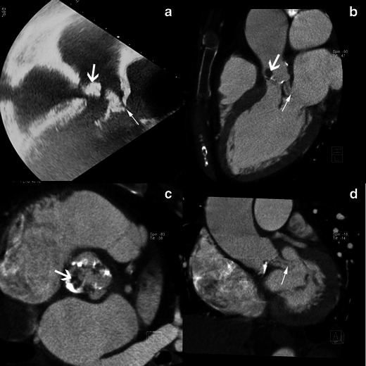
Fig. 6.

Results of echocardiography (TEE) and MSCT studies in a case of aortic valve infective endocarditis complicated by a fistula between aorta and left atrium. The TEE study is shown in the 120-degree long-axis view (a) and the MSCT acquisition with MPR reconstructions in the LVOT view (b), the aortic valve plane view (c), the coronal oblique view at the level of the aortic root (d) show a fistula between aortic root and the left atrium (thin white arrow) and a large vegetation on the aortic valve (thick white arrow)
